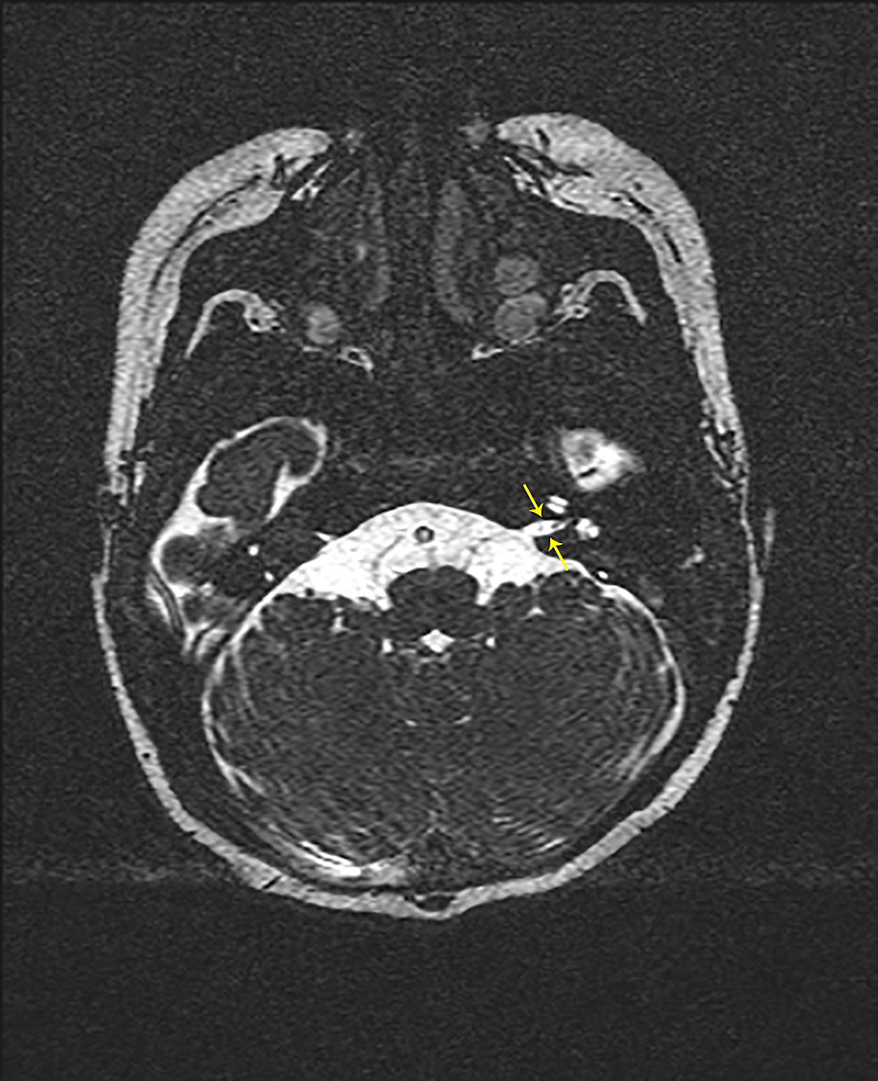
Herein, we outline the most useful MRI sequences, review the imaging characteristics of the most frequently encountered pathologic entities, and describe the use of MRI in the postoperative follow-up of vestibular schwannomas and cholesteatomas. Radiologists who interpret such imaging studies should be well-versed in the expected appearances of the most common pathologic entities. In many cases, MRI is complementary to CT in the evaluation of surgical candidates, such as individuals being considered for pediatric cochlear implantation, where the labyrinth may be malformed and the facial nerve may take an anomalous course through the temporal bone. Common applications of MRI include diagnostic evaluation of sensorineural hearing loss, assessment of cochlear implant candidacy, monitoring for residual or recurrent cholesteatoma within the tympanomastoid space, and monitoring for vestibular schwannoma within the inner auditory canal or cerebellopontine angle. MRI of the temporal bone and lateral skull base has become a mainstay of neuroradiology to delineate anatomic structures and to investigate neoplastic or infectious processes affecting this complex space. For this journal-based CME activity, author disclosures are listed at the end of this article. The ACCME requires that the RSNA, as an accredited provider of CME, obtain signed disclosure statements from the authors, editors, and reviewers for this activity. Physicians should claim only the credit commensurate with the extent of their participation in the activity. The RSNA designates this journal-based SA-CME activity for a maximum of 1.0 AMA PRA Category 1 Credit ™. The RSNA is accredited by the Accreditation Council for Continuing Medical Education (ACCME) to provide continuing medical education for physicians. ■ Describe the typical MRI appearance of acute labyrinthitis and labyrinthitis ossificans ■ Describe the poor prognostic imaging features of vestibular schwannomas ■ Identify the major anatomic structures visible on MRI within the internal auditory canal (IAC), labyrinth, and middle ear In addition, the features at pre- and postprocedural MRI will be discussed to help ensure that diagnostic radiologists may be of greatest use to the ordering physicians.Īfter reading the article and taking the test, the reader will be able to:


The purpose of this review is to provide an overview of the most useful MRI sequences for internal auditory canal and labyrinthine imaging, review the relevant anatomy, and discuss the expected appearances of the most commonly encountered pathologic entities.

Nevertheless, despite the widespread use of MRI for these purposes, many radiologists remain unfamiliar with the complex anatomy and expected imaging findings with such examinations. It is also extensively used in pre- and postoperative evaluations, particularly in patients with vestibular schwannomas and candidates for cochlear implantation. It is used to evaluate normal anatomic structures, evaluate for vestibular schwannomas, assess for inflammatory and/or infectious processes, and detect residual and/or recurrent cholesteatoma. MRI is firmly established as an essential modality in the imaging of the temporal bone and lateral skull base.


 0 kommentar(er)
